
- Visual Field Test for Blepharoplasty
- What Is the Test for Eyelid Surgery?
- The Procedure of Visual Field Test for Blepharoplasty
- The First Step
- The Second Step
- The Importance of Visual Field Test for Blepharoplasty
- Conditions That Can Affect the Visual Field
- Cataracts
- Eye Tumors
- Glaucoma
- Pituitary Gland Disorders
- Cerebrovascular Accident
- Retinal Detachment
- Multiple Sclerosis
- Retinal Hemorrhage
- Benign and Malignant Brain Tumors
- Brain Abscess
- Macular Degeneration
- Types of Visual Field Tests
- Automated Static Perimetry Test
- Kinetic Visual Field Test
- Frequency Doubling Perimetry
- Electroretinography
- Amsler Grid
- Confrontational Visual Field Exam
- Other Types of Eye Tests
- Refractive Test
- Visual Acuity Test
- Eye Muscles Test
- Retinal Examination
- Glaucoma Testing
- Color Blind Test
- The Bottom Line
A visual field test assesses the integrity and health of vision, which helps detect physiological dysfunctions. Some doctors suggest a visual field test for blepharoplasty, which is a cosmetic surgery to improve the shape of your eyelid.
Many methods are available to test the visual field. Some of these methods are simple compared to more advanced and technical methods. However, people who plan to undergo blepharoplasty are sometimes asked to get a special visual test called the "Ptosis visual field test".
Ptosis refers to the drooping of the upper eyelid over the eye. This issue is troublesome and a source of great dissatisfaction among people. Blepharoplasty or eyelid surgery can address ptosis and bring relief to those people.
Visual Field Test for Blepharoplasty
Most clinics use a visual field to prove the presence of ptosis and justify blepharoplasty, but there is no standardized visual field designed for this exact purpose. Clinics often use either Goldmann visual field test or Humphrey visual field test to diagnose different eye conditions.
Although Humphrey visual field test is commonly used to assess ptosis, it isn always reliable to measure the extent of ptosis before blepharoplasty. Thus, in recent years, researchers tried to develop a special visual field test that can be used to assess ptosis and make the case for blepharoplasty. They called it a "Ptosis Visual Field Test", and is designed to assess the impact of drooping eyelids on vision.
Ptosis visual field test will not just assess ptosis, but also other potential problems in the eye. The test results help doctors recognize certain types of injury or disease and track the changes that occur before and after eyelid surgery.
What Is the Test for Eyelid Surgery?
According to experts, the Ptosis Visual Field Test could be the best test for eyelid surgery as it explores the degree to which your eyelids obstruct your vision. The same test can be used as a field vision test for droopy eyelids.
Both the left and right eyes will each be tested twice during the examination. First, the eyelids will be in their typical drooping posture. Next, they will tape them gently with tape that is safe for the skin around the brow and eyelids. Finally, they will retest to see how much your vision has improved.
Often, it takes around 20 minutes to complete the test. Asking for lubricating drops or a break during the test is normal. If a patient has ptosis of the eyelid you can expect to see the eyelid having a droopy appearance or eyelid overhang.
The Procedure of Visual Field Test for Blepharoplasty
Ptosis visual field test usually takes around 20 minutes, and your doctor will assess both of your eyes. This test can be administered in two ways: taped and untaped. Visual field test taped and untaped is determined based on the needs of the patient. Doctors will perform two steps to assess each eye:
The First Step
Your doctor will examine your eyelids in their normal droopy position without modifying their position. The goal is to measure your vision ability without lifting your eyebrow.
The Second Step
Your doctor will examine your eyelids after taping them up with a gentle tape and retest your vision again to measure any difference. This can give a fairly accurate idea about what your vision would be like without the droopy skin in the way.
The Importance of Visual Field Test for Blepharoplasty
The optical or visual field is the maximum visual capacity a person can have in the eye center and on the sides without moving their eyes. In other words, the visual field is seeing a thing and its surroundings.
When you drive a car, for example, you need to see the road and check the mirror and the front view at the same time. You can do that through the visual field you have. The same thing is true for mothers and those who work in security jobs. They need to see the complete visual field to accomplish their tasks.
If the visual field test indicates a problem in vision due to droopy eyelids, eyelid surgery or blepharoplasty could be the key to solving the problem. But it doesn end here!
In fact, a visual field test should be repeated more than one time if the patient is tired because being just tired can affect the results of the test. Having droopy eyelids makes you look tired all the time.
Conditions That Can Affect the Visual Field
You can have a visual field test for blepharoplasty, but this test also can detect other diseases and conditions that can cause problems for the visual field, including:
Cataracts
The general sign of cataracts is the presence of cloudiness in the lens of the eye. This leads to less light entering the eyes, an inability to distinguish soft colors, as well as blurred vision. Patients feel as if they see from behind a curtain as the images become blurry. This leads to limitations in the visual field.
Among the main risk factors of cataracts are aging, taking some medications, diabetes, prolonged exposure to the sun, as well as genetic factors, shocks, and accidents. Doctors commonly suggest cataract surgery with phacoemulsification to fix the problem.
Eye Tumors
Eye tumors can lead to a limited visual field as they affect one or more parts of the eye. These tumors may be primary; meaning they originate in the eye itself, or metastatic; meaning they are formed in other parts of the body, such as the kidney, breast, lung, prostate, or thyroid gland. Metastatic tumors spread to the eye in the later stages of cancer.
The major types of eye tumors are eyelid tumors, eye socket tumors, retinal tumors, and conjunctival tumors. These tumors may be benign or malignant.
There are various methods to diagnose eye tumors, including biopsy, fluorescein angiography, and ultrasound imaging.
Glaucoma
Glaucoma is one of the most common conditions that lead to visual field impairments. Most often this condition affects the eyes due to increased intraocular pressure. Unfortunately, this condition is one of the most common conditions that can lead to vision loss in people over sixty years old.
Symptoms of glaucoma appear gradually and include the appearance of halos around the light, eye pain, headache, and blurred vision. Glaucoma is linked to optic nerve damage, which causes dark spots to appear in the field of vision.
Scientists also linked this condition with a high level of intraocular pressure resulting from fluid accumulation. Genetic factors play an important role in developing the condition. To diagnose glaucoma, your doctor can perform a glaucoma visual field test to examine your visual field. Glaucoma surgery is available to treat the condition.
Pituitary Gland Disorders
Pituitary gland disorders lead to a disruption in the secretion of many hormones necessary for multiple functions in the body, including growth hormone, adrenal gland-stimulating hormone, milk-stimulating hormone (prolactin), luteinizing hormone, thyroid-stimulating hormone, and antidiuretic hormone. One of the symptoms of pituitary gland disorders is visual field impairment.
Cerebrovascular Accident
Cerebrovascular accident or stroke occurs because of a lack of oxidative blood flow to parts of the brain, which causes brain cell death. Among the causes of stroke are high blood pressure, obesity, smoking, diabetes, and atrial fibrillation.
Among the symptoms of a stroke are sudden headache, slurred speech, problems in the field of vision, numbness or tingling in the face, leg, or arm, difficulty walking, and imbalance.
Retinal Detachment
Retinal detachment is an urgent condition that leads to severe impairment in the visual field. It occurs as a result of the displacement of part of the retinal tissues in the back of the eye, which leads to the separation of the retinal wall from the blood vessels that supply cells with oxygenated blood and other nutrients. If retinal detachment persists without treatment, this may cause vision loss.
Among the main signs that predict the occurrence of a retinal detachment are the appearance of flashes and blurring in the peripheral vision and the appearance of a shadow that obscures vision.
Multiple Sclerosis
Multiple sclerosis affects the outer shell surrounding the nerve cells in the brain and spinal cord. This causes communication disturbance between the brain and the rest of the body. Visual field impairment or total loss of vision (mainly in one eye) is one of the major symptoms of the disease. Other symptoms include numbness in the extremities, persistent fatigue, speech difficulties, sexual dysfunction, and tingling sensations in various parts of the body.
Retinal Hemorrhage
The retina is a thin layer consisting of light-sensitive tissues located at the back of the eye. Retinal hemorrhages often occur due to a disturbance in the blood sugar level, high blood pressure, or blockage of eye blood vessels. This causes various complications, including visual field impairment.
Benign and Malignant Brain Tumors
Brain tumors are abnormal growth of cells in the brain. These tumors could be benign or malignant. The tumor may arise primarily in the brain, or it may arise secondarily as a result of migrating tumors from other places in the body.
The major types of brain tumors include but aren limited to choroid plexus carcinoma, acoustic neuroma, craniopharyngioma, meningioma, neuroblastoma, oligodendroglioma, and astrocytoma.
Brain Abscess
A brain abscess occurs primarily in the brain tissues due to infection with germs that reach the brain through contaminated blood or through teeth, nose, or ears. The symptoms of this disease are diverse and may include high body temperature, inability to walk, weakness in the limbs, speech problems, and blurred vision.
Macular Degeneration
Macular degeneration is among the common eye conditions that can cause visual field impairment. This condition often occurs in people over the age of fifty.
It leads to blurry vision and impairment in central vision. The reason for this stems from the decreased size of the macula, which is located behind the retina and helps you read things, see detailed images, and distinguish colors.
The major symptoms of macular degeneration include difficulty perceiving images, blurred vision, and inability to distinguish images. The condition can progress to a more serious form because of an aneurysm in the blood vessels that supply the retina, leading to vision deterioration. A macular degeneration surgery is available to deal with the condition.
Types of Visual Field Tests
There are at least 6 types of visual field tests. We can mention them as follows:
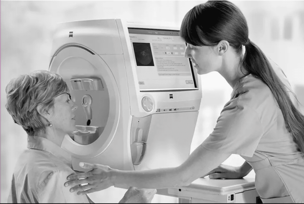
Automated Static Perimetry Test
This test can create a detailed map of how you see objects in your field of vision. You will set a front of a “perimeter” and look at a center target. You will press a button when seeing a blinking light. Your position will be fixed, so your doctor will determine which lights you see outside of your central area of vision. The visual field test for blepharoplasty commonly depends on this method.
Kinetic Visual Field Test
Instead of seeing a blinking light like in the previous test, you will see a moving light during this test. Hence, the difference between a kinetic visual field test and an automated static perimetry test is minor.
Frequency Doubling Perimetry
Through this test, your doctor will be able to assess your visual field by utilizing optical illusions that flicker at varying rates. These optical illusions come in the form of vertical black-and-white bars. The inability to see these bars at certain points in the test could indicate a vision problem in the field of vision.
Electroretinography
Electroretinography assesses visual field loss resulting from retinal conditions. Eyes dilation is a part of this test, and they should be kept open throughout the test. Electroretinography measures the electrical signals of cells in the retina (photoreceptors).
Amsler Grid
This test is among the simplest tests used by doctors to examine the visual field. . All you must do is look at a dot in the middle of a grid. Some forms of Amsler grid can be used at home to look for age-related problems that can affect the visual field.
Confrontational Visual Field Exam
This might be the simplest test in the list. During this test, the doctor faces the patient at a distance of one meter and asks them to cover one eye and see through the other eye only. The doctor moves their fingers to the area outside the visual field of the examinee and asks them to tell what they e seeing.
Other Types of Eye Tests
There are various eye tests other than visual field tests, including the following:
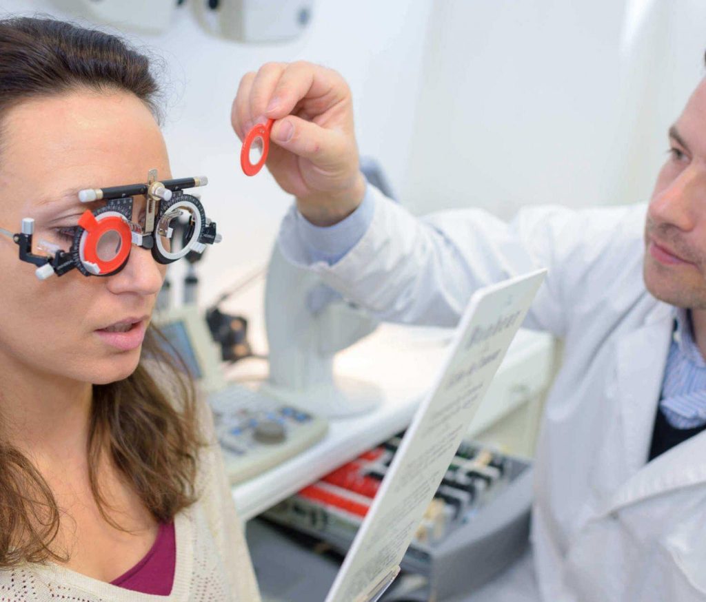
Refractive Test
This test measures the extent to which light is directed towards the inside of the eye pupil. If refraction is absent, this may require correction using eyeglasses, refractive surgery (LASIK), or contact lenses to restore vision. This test helps determine the exact specifications of the required lenses.
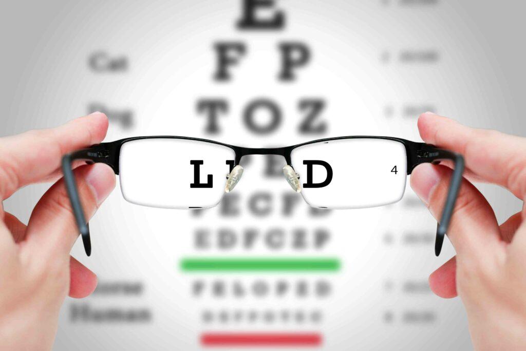
Visual Acuity Test
This test measures the extent of visual acuity and clarity of vision. The doctor could use a Snellen chart, which includes a group of alphabet letters printed on a board and displayed at a distance from the patient. The size of the letters varies from largest to smallest. Each eye is examined separately, and the patient must determine every letter.
Eye Muscles Test
Doctors rely on this test to evaluate the control and coordination of eye muscles. The doctor assesses the movement of the eye while the eye follows one of the moving objects, and then diagnoses the patients condition based on that.
Retinal Examination
This examination helps discover problems and disorders in the back of the eye. Doctors also call it “ophthalmoscopy” and often choose it to evaluate the rear parts of the eye, including the retina and blood vessels. Doctors use eye drops to dilate the pupil to make it easier to see more accurately. The examination of the retina is divided into:
- Direct ophthalmoscopy: The doctor uses an ophthalmoscope and directs light toward the pupil to be able to see the rear parts.
- Indirect ophthalmoscopy: The doctor wears a headband with a lens. They also use a bright light to help see the internal structure of the eye.
Glaucoma Testing
Glaucoma testing aims at identifying the level of fluid pressure inside the eye to detect glaucoma that causes nerve damage within the eye. Prominent testing methods include:
- Applanation tonometry: This is one of the most common methods to examine eye pressure. It involves the use of a slit lamp. The doctor moves the device over the cornea and measures the level of pressure inside the eye.
- Non-Contact tonometry: The doctor directs air toward the eye to measure intraocular pressure without touching any parts of the eye. This does not require anesthesia as the patient only feels an instantaneous spurt of air.
Color Blind Test
The color blind test evaluates your ability to distinguish between colors. The specialist doctor displays a set of different points with multiple colors. If no problem in color vision is present, the patient is supposed to identify the colors accurately. On the contrary, if the patient finds it difficult to distinguish between colors, this hints at the presence of a color recognition problem.
The Bottom Line
A visual field test for blepharoplasty or "Ptosis visual field test" is not always necessary before surgery, but some surgeons suggest it in certain circumstances. It can help them assess the droopy eyes and their impact on your vision.
Blepharoplasty visual field testing is different than other eye tests, such as visual acuity tests and ophthalmoscopy. But, if your plastic surgeon finds it necessary to check your eye and screen eye diseases before blepharoplasty, they may suggest one of the other sophisticated eye tests.
At International Clinics, a visual field test for blepharoplasty is available for patients who suffer from an extreme case of droopy eyes. You can reach us directly by using the Contact Us buttons below.
Read more:
How to Qualify for Eyelid Surgery?
Upper Blepharoplasty Cost: Affordable Eyelid Surgery