
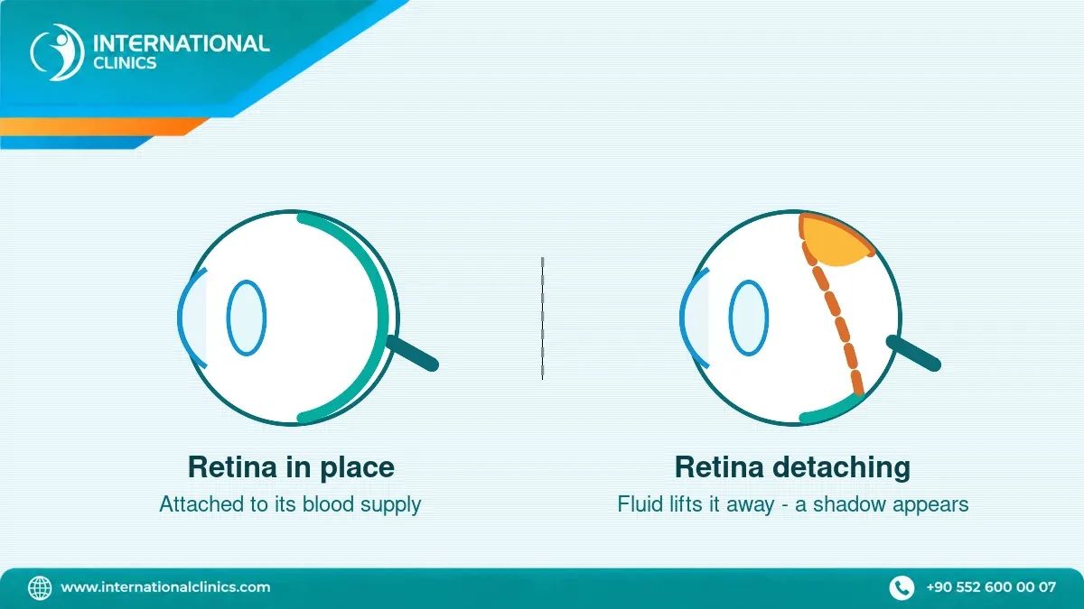
Retinal detachment symptoms are a sudden shower of floaters, flashes of light, a dark curtain or shadow moving across your sight, blurred or distorted vision, and loss of side vision. The detachment itself is painless, which is exactly why it is missed — and it is a same-day emergency.
Retinal Detachment Symptoms: The Five Warning Signs
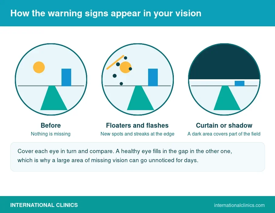
Five warning signs matter, and the table below is the reference list used throughout this page. Any one of them, appearing suddenly, is a reason to be seen the same day by an eye specialist rather than to wait and see whether it settles.
| Warning sign | What people actually describe | How urgent |
|---|---|---|
| Sudden floaters | A shower or sharp increase in small dark spots, squiggly lines or cobwebs drifting across vision | Same-day assessment |
| Flashes of light | Brief streaks of lightning or flickering, usually at the edge of vision and often worse in the dark | Same-day assessment |
| A dark curtain or shadow | A grey veil or curtain moving in from one side, the top or the bottom of the visual field | Emergency — the retina is already detaching |
| Blurred or distorted vision | Reduced sharpness, or straight lines such as door frames appearing wavy or bent | Emergency if it starts suddenly |
| Loss of side vision | A gradual or sudden loss of peripheral sight, as though part of the picture has been cut away | Emergency — central vision may be next |
These five are the signs listed by the NHS and Mayo Clinic, and they are the same five that Google’s own summary of this question puts first. The detachment is not always complete when they begin: the earliest stage is often a retinal tear, which is far easier to treat.
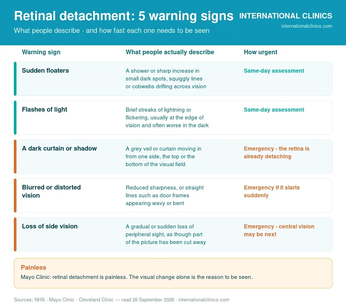
Every item in this infographic is taken from the warning-sign table above.
Are Eye Floaters and Flashes Always a Warning Sign?
No. Floaters are extremely common and most are harmless — small clumps in the vitreous gel casting shadows on the retina. What matters is change. A few floaters you have had for years are not the warning sign; a sudden shower of new ones is, especially when flashes appear at the same time.
The combination is the alarm: new floaters plus flashes of light means the vitreous is pulling on the retina, and that pull is what tears it. The NHS advice is to contact NHS 111 when floaters suddenly appear or suddenly increase in number, when you see flashes, or when a shadow or sudden blurring appears. If you are unsure whether what you see has changed, that uncertainty is itself a reason to be examined.
Retinal Tear Symptoms vs a Full Retinal Detachment
A retinal tear is a hole in the retina; a retinal detachment is the retina lifting away from the wall of the eye once fluid has passed through that hole. The symptoms overlap almost completely at the start, which is why the two cannot be told apart at home.
| Retinal tear | Retinal detachment | |
|---|---|---|
| Typical symptoms | New floaters and flashes; vision usually still complete | The same, plus a shadow, curtain or missing area of vision |
| What has happened | The retina is torn but still in place | Fluid has lifted the retina off its blood supply |
| Usual treatment | Laser or freezing treatment, normally in the clinic | Surgery in an operating theatre |
| Why the difference matters | Sealing a tear can prevent the detachment entirely | Vision already lost may not fully return |
This is the practical argument for going in on the day the flashes and floaters start rather than the week after: the same eye, seen earlier, is often a clinic laser appointment instead of theatre surgery. Diagnosis is made by examining the back of the eye with the pupil dilated, supported where needed by retinal imaging.
Is Retinal Detachment Painful?
No, and this is the most dangerous single fact about it. Mayo Clinic states it plainly:
“Retinal detachment is painless. But warning signs almost always appear before it occurs or has advanced… See a healthcare professional right away if you have any symptoms of retinal detachment. This condition is an emergency that can cause lasting vision loss.”
— Mayo Clinic, Retinal detachment: symptoms and causes (read 26 September 2026)
Because there is no pain, people apply the wrong test. Pain is what usually sends someone to a doctor, so a painless change in vision gets filed as tiredness, a migraine or a smudge on a lens. The trigger to seek help has to be the visual change alone. A painless eye is not a reassuring eye.
How Long Before Retinal Detachment Causes Blindness?
There is no single clock that fits every eye, and any source that gives you one exact number is guessing. What is consistent across the evidence is the direction: the longer the retina stays detached, the less vision returns, and the part of the retina that is affected decides how fast the damage matters.
- While central vision is still normal — the centre of the retina, the macula, is still attached. This is the best position to be operated on from, and it is measured in days, not weeks.
- Once a curtain covers the centre of vision — the macula has detached. Surgery can still reattach the retina, but sharpness that was lost before the operation may not fully come back.
- Weeks of delay — scar tissue forms on the retina, the repair becomes harder, and more than one operation is more likely.
So the honest answer to how long you have is: nobody can tell you, and the question itself is the wrong one. Treat the first shadow as today’s problem. The same reasoning applies to the other eye conditions that steal sight quietly — see glaucoma symptoms and cataract symptoms for how differently each one announces itself.
Who Is Most at Risk of a Detached Retina?
Retinal detachment is not common in absolute terms. A 2026 global review in Epidemiologia puts the annual incidence of the most frequent form, rhegmatogenous retinal detachment, at 12.17 per 100,000 people. But that average hides very large differences between people, and the risk factors below are the reason.
| Risk factor | What the evidence shows | Source |
|---|---|---|
| Age | More common between 40 and 70; incidence peaks at 60–70 | Mayo Clinic; Epidemiologia 2026 |
| Short-sightedness (myopia) | Mild myopia under 3 dioptres carries about a 3-fold risk; high myopia over 6 dioptres up to 39-fold | Epidemiologia 2026 |
| Previous cataract surgery | Raises the risk 4–10-fold, with a cumulative incidence of 1–2% within 5–10 years | Epidemiologia 2026 |
| A previous detachment | Detachment in one eye raises the risk in the other | Mayo Clinic |
| Family history | Listed as a risk factor in its own right | NHS; Mayo Clinic |
| Severe eye injury or past eye surgery | Both are listed risk factors | NHS; Mayo Clinic |
| Poorly controlled diabetes | Scar tissue from diabetic eye disease can pull the retina away | Mayo Clinic |
The myopia figures are the ones worth reading twice. If you are strongly short-sighted, floaters and flashes are not a minor complaint to mention at your next routine test — what causes short-sightedness explains why the shape of a myopic eye puts the retina under strain in the first place. If your risk comes from diabetes instead, the mechanism is different: scar tissue on the retina, not a tear in it.
What Are the Three Types of Retinal Detachment?
Mayo Clinic groups retinal detachment into three types. They share the same warning signs but not the same cause, and the cause decides the operation.
| Type | What causes it | Who it typically affects |
|---|---|---|
| Rhegmatogenous | A hole or tear lets fluid collect underneath the retina and lift it away | The most common type; linked to ageing of the vitreous and to myopia |
| Tractional | Scar tissue on the surface of the retina contracts and pulls it off | Most often people with poorly controlled diabetes |
| Exudative | Fluid builds up behind the retina with no tear at all | Linked to macular degeneration, injury, inflammation, infection or tumours |
The exudative type is the reason a full examination matters rather than a quick look: it is treated by treating the condition behind it, not by sealing a hole that is not there. Where the underlying problem sits at the centre of the retina instead, blurred and distorted central vision is the presenting complaint rather than a curtain at the edge.
Retinal Detachment Surgery Options and What They Cost
Retinal detachment surgery has one aim: put the retina back against the wall of the eye and seal whatever let the fluid through. Which operation is used depends on the type, the size and where the tear sits. The prices below are our published figures for retinal detachment treatment in Turkey.
| Procedure | What it involves | Our published price |
|---|---|---|
| Laser treatment or cryopexy | Heat or freezing seals a tear before the retina lifts; usually done in the clinic | $500–$1,000 |
| Pneumatic retinopexy | A gas bubble is injected to press the retina back into place | From $1,500 |
| Scleral buckling | A silicone band is placed around the eye to relieve the pull on the retina | $1,800–$2,800 |
| Vitrectomy | The vitreous gel is removed and replaced with gas or oil so the retina can settle | $2,500–$3,500 |
| Combined or advanced procedures | For example vitrectomy with silicone oil for long-term stabilisation | May exceed $4,000 |
Across procedures the range is $1,500 to $3,500 per eye, and the figure depends on the technique, the case and the facility. Tears caught early sit at the bottom of that table, which is the financial version of the same argument the clinical evidence makes. Laser therapy for retinal diseases explains the laser pathway in more detail, and the full breakdown lives on our retinal detachment treatment page.
Things to Avoid With Retinal Detachment
Between noticing symptoms and being examined, the sensible list is short:
- Do not wait for the symptoms to settle. A shadow that fades is not proof the retina is safe.
- Avoid heavy lifting, straining and vigorous exercise until an eye specialist has looked at the retina.
- Do not fly before you have been assessed, and never after gas has been placed in the eye until your surgeon says the bubble has gone — cabin pressure expands it.
- Do not drive if part of your visual field is missing.
- Do not rely on the other eye to tell you how bad it is. Cover each eye in turn and compare; a good eye hides a large gap in the other.
After surgery the restrictions are set by your surgeon and depend on the operation. Where a gas bubble is used, position matters: the NHS notes that you may be asked to keep your head in a particular position for up to 7 days.
Recovery After Retinal Detachment Surgery
Recovery is slower than most people expect, and it is normal to see poorly for a while after a technically successful operation. According to the NHS, vision is usually blurred for 2 to 6 weeks after surgery, and a specific head position may be required for up to 7 days when gas is used inside the eye.
Two expectations are worth setting honestly. First, reattaching the retina and restoring sharp sight are not the same achievement — how much vision returns depends heavily on whether the centre of the retina had already detached. Second, more than one operation is sometimes needed, and that is a recognised part of the pathway rather than a sign something went wrong.
Risks, Limits and When to Treat Symptoms as an Emergency
Retinal detachment surgery carries real risks, and no honest page about the symptoms can end without them. Recognised risks include bleeding inside the eye, infection, raised pressure, cataract formation after vitrectomy, further detachment requiring another operation, and incomplete recovery of vision. Outcomes vary from person to person and no result can be promised in advance.
Treat the following as a medical emergency and seek care the same day:
- A sudden shower of new floaters, with or without flashes of light
- Flashes of light that are new, repeated, or worse in a dark room
- A dark curtain, veil or shadow moving across any part of your vision
- A sudden loss of side vision, or straight lines that have started to bend
- Any sudden painless change in vision after an eye injury or eye surgery
In the UK, NHS advice is to contact NHS 111 for these symptoms; elsewhere, go to an eye casualty or emergency department. This page is general information and does not replace an examination. If you would like your symptoms and scans reviewed by our ophthalmology department, our patient team can arrange it — and if you already have a diagnosis, our retinal detachment treatment guide sets out the options and costs.