The retina is a thin layer located at the back of the eye, responsible for receiving light and converting it into electrical signals that are transmitted to the brain via the optic nerve to form a visual image.
It is richly supplied with blood vessels that nourish its tissues, and any damage to the retina, its blood vessels, or the optic nerve can lead to permanent vision loss.
Retinal imaging tests are essential for evaluating retinal health and detecting early signs of damage.
This article explains retinal imaging tests in Turkey, including their types, techniques, and the importance of undergoing these tests.
For more details and a personalized cost estimate, contact International Clinics and book a free consultation.
CONTENT_TABLE_AREA
What is Retina?
The retina is the part of your eye that collects the eye and converts it to an electric signal that can be sent by the optic nerve to the brain to be transparent into a picture.
The retina is a light-sensitive, thin layer of tissue consisting of light-sensitive cells that line the back of the eyeball.

What is A Retina Imaging Test?
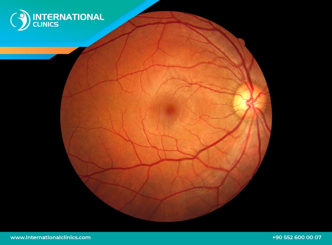
Retinal imaging is a diagnostic test that captures high-resolution digital images of the inner, back surface of the eye. These images provide a clear view of the retina, optic nerve, and blood vessels that supply the eye’s tissues.
This technology allows eye doctors to examine more than 80% of the retina in a single capture, aiding in the early detection of eye conditions. The test is often performed alongside routine eye exams and pupil dilation to assess retinal health and detect any damage caused by primary eye diseases or systemic conditions.
What does retinal imaging show?
1. Overgrowth or damage in blood vessels: Indicates diabetic retinopathy or retinal vascular occlusion.
2. Retinal destruction or degeneration: This may be a sign of age-related macular degeneration, retinal detachment, or retinal inflammation.
3. Optic nerve damage: Suggests glaucoma or optic neuritis.
Who Needs a Retinal Imaging Test?
-
People experiencing blurry vision, floaters, or difficulty seeing.
-
Patients with chronic diseases that may affect the retina and vision, such as diabetes mellitus or hypertension.
-
Elderly individuals who have a higher risk of eye conditions.
-
Patients with progressive vision loss.
-
Patients at high risk of developing diabetic retinopathy, macular degeneration, ocular melanoma, or glaucoma.
Medical Conditions That Require Retinal Imaging Tests
Common diseases can be detected by retina imaging
Diabetic retinopathy: uncontrolled diabetes mellitus associated with a high risk of vision loss, that is because frequently elevated blood sugar damages eye blood vessels.
Diabetic patients suffering from blurred vision or vision deterioration have to check their eye blood vessels with a retina imaging test
Macular degeneration:
This condition is an age-related condition, in which the macula, the center part of the retina, starts to damage and lose functionality, this occurs due to abnormal blood growth under the retina. The blood loss in macular degenerative disease is fast and rapid medical intervention is crucial. Retinal imaging is a very helpful tool to detect macular degeneration.
Glaucoma: is the damage of the optic nerve that results from severe or chronic elevation in the intraocular pressure due to fluid accumulation in the front of the eye.
Besides the other medical tests for eye pressure measurements, glaucoma retinal image is a helpful tool to detect any optic nerve damage early, this is crucial to manage the glaucoma properly by choosing the best effective glaucoma treatment, such as glaucoma surgery.

Types of Retinal Imaging Tests
Several Tests Help Examine the Retina and Detect Potential Abnormalities:
1. Optic Nerve Examination:
Techniques like fundoscopy (ophthalmoscopy) use a special light to directly examine the retina and optic nerve, helping detect conditions such as optic neuropathy or glaucoma.
2. Retinal Imaging Tests:
These tests capture images of the retina for closer evaluation.
- Fundus Photography provides detailed images for documentation and monitoring of retinal health.
- Optical Coherence Tomography (OCT) offers high-resolution, cross-sectional images, aiding in the diagnosis of conditions like macular edema and retinal detachment.
3. Blood Vessel Assessment:
This can be done using techniques with or without dye.
- Fluorescein Angiography (FA) involves injecting a fluorescent dye to highlight blood flow in the retina, useful for detecting diabetic retinopathy and macular degeneration.
- Non-dye techniques like OCT Angiography (OCTA) allow visualization of blood vessels without contrast injection.

Retinal Imaging Technology
Types of retina imaging tests involve both traditional fundus test techniques and other advanced technologies.
Advanced technology in retinal imaging helps in the early detection of eye diseases, allows for more precise diagnosis, and is better at monitoring disease progression over time.
1. Fundus Photography or Retinal Fundus Imaging
Fundus Photography is ordered to document the status of the optic nerve, macula, retina, blood vessels, and the vitreous.
Several fundus tests are performed as part of retinal imaging, each serving a different purpose.
Not all tests are required for every patient, doctors choose the most suitable one based on the suspected condition.
- Color Fundus Photography: Captures high-resolution color images of the retina.
- Red-Free Fundus Photography: Enhances contrast by removing red light, useful for examining blood vessels.
- Fundus Autofluorescence (FAF): Detects metabolic changes in retinal pigment epithelium (RPE) without using dye.
2. Angiographic Imaging
These techniques use contrast dyes to visualize blood flow and detect abnormalities:
- Fluorescein Angiography (FA): Uses fluorescein dye to assess retinal blood vessels and circulation.
- Indocyanine Green Angiography (ICGA): Provides deeper visualization of choroidal blood vessels.
3️. Advanced Retinal Imaging
More detailed imaging techniques that provide in-depth analysis of the retina:
- Optical coherence tomography (OCT): Creates high-resolution cross-sectional images of the retina.
- OCT Angiography (OCTA): Non-invasive visualization of blood flow in retinal vessels without dye.
- Optomap- ultra-widefield retinal imaging: Captures up to 200° of the retina in a single image
4️. Direct Retinal Examination (Ophthalmoscopy)
Traditional methods used for real-time examination of the retina:
- Direct Ophthalmoscopy: Provides a magnified view of the central retina.
- Indirect Ophthalmoscopy: Uses a hand-held lens for a wider view of the peripheral retina.

Why Choose Turkey for A Retinal Image Test?
Experienced retina specialists:
A retina specialist is an ophthalmologist with advanced training in diagnosing, medically managing, and surgically treating diseases affecting the retina, macula, and vitreous.
They specialize in conditions such as diabetic retinopathy, macular degeneration, retinal detachment, and retinal vascular diseases.
Turkey is home to highly skilled retina specialists and ophthalmologists trained in top international institutions.
Many doctors have extensive experience in diagnosing and treating complex retinal conditions, ensuring high-quality care.
Advanced Retinal Imaging Technology
Hospitals and eye clinics in Turkey are equipped with the latest retinal imaging technologies, including Fundus Photography for detailed retinal evaluation, Optical Coherence Tomography (OCT) for high-resolution cross-sectional imaging, Fluorescein Angiography (FA) & Indocyanine Green Angiography (ICGA) for assessing retinal blood flow, and Ultra-Widefield Retinal Imaging (Optomap) for capturing a larger view of the retina.
Affordable retina imaging costs:
Turkey offers cost-effective medical services compared to many Western countries, without compromising quality.
Retina imaging procedures in Turkey can be 50-70% cheaper than in the US or Europe.
The average cost of common retina tests in Turkey:
- Fundus Photography: $50 - $150
- Optical Coherence Tomography (OCT): $100 - $300
- Fluorescein Angiography (FA): $150 - $400
Other advanced common eye procedures that are effectively and successfully performed in Turkish clinics include
LASIK Eye Surgery in Turkey 2025 and Phacoemulsification in Turkey, the advanced method to treat cataracts safely and effectively.
FAQs
What does retinal imaging test for?
Retinal imaging tests for abnormalities in the retina, optic nerve, and blood vessels, helping detect conditions like diabetic retinopathy, macular degeneration, glaucoma, and retinal detachment.
What is the retinal imaging procedure?
The procedure involves capturing high-resolution images of the retina using specialized imaging techniques such as fundus photography, OCT, or fluorescein angiography, sometimes with pupil dilation for a clearer view.
What is the difference between an OCT scan and retinal imaging?
OCT is a specific type of retinal imaging that provides cross-sectional images of the retina, while general retinal imaging includes various techniques like fundus photography and angiography for broader retinal assessment.
Is retinal imaging better than dilation?
Retinal imaging provides detailed digital images for documentation and diagnosis, while dilation allows for a direct, real-time eye exam. Both are useful, and doctors may recommend one or both depending on the case.
What is retinal imaging called?
Retinal imaging is also known as fundus photography, retinal photography, or optical coherence tomography (OCT), depending on the technique used.
Is retinal imaging worth it?
Yes, retinal imaging is valuable for early detection of eye diseases like diabetic retinopathy, macular degeneration, and glaucoma, helping prevent vision loss with timely treatment.
What is the difference between OCT and retinal imaging?
OCT is a type of retinal imaging that provides cross-sectional views of the retina, while general retinal imaging includes other techniques like fundus photography and fluorescein angiography.
Is retinal imaging necessary?
Retinal imaging is not always necessary but can be useful for detecting eye diseases early, especially if you have risk factors like diabetes or a family history of eye conditions.
Should I get retinal imaging?
If you have vision concerns, high risk of eye disease, or it's recommended by your eye doctor, getting retinal imaging can be beneficial.
What is retinal imaging?
Retinal imaging is a diagnostic technique that captures detailed images of the back of your eye, including the retina, optic nerve, and blood vessels.
What can retinal imaging detect?
It can detect conditions like diabetic retinopathy, glaucoma, macular degeneration, retinal detachment, and even signs of systemic diseases like hypertension.
Do I need retinal imaging every year?
It depends on your eye health; people with risk factors or existing conditions may need it annually, while others might only need it occasionally.