Retinal imaging is an important way to diagnose many problems that occur in the eye. However, many people remain unaware of the difference between Optomap retinal imaging vs dilation.
Advanced medical research and numerous studies have shown that retinal imaging is a safe way to diagnose problems in the eye. Eye dilation is also needed to diagnose and check eye health in certain cases.
Retinal imaging is a diagnostic procedure that depends on photography. It aims at obtaining multiple images of the eye fundus and retina to check and detect various diseases that affect the retina, such as macular degeneration, diabetic retinopathy, or retinitis pigmentosa.
Thankfully, retinal imaging is very safe and harmless to the eye and does not cause any problems. But to better understand retinal imaging, it is necessary to explore the difference between retinal imaging and dilation. This is what we reveal through the following lines.
Optomap Retinal Imaging vs Dilation
Dilation
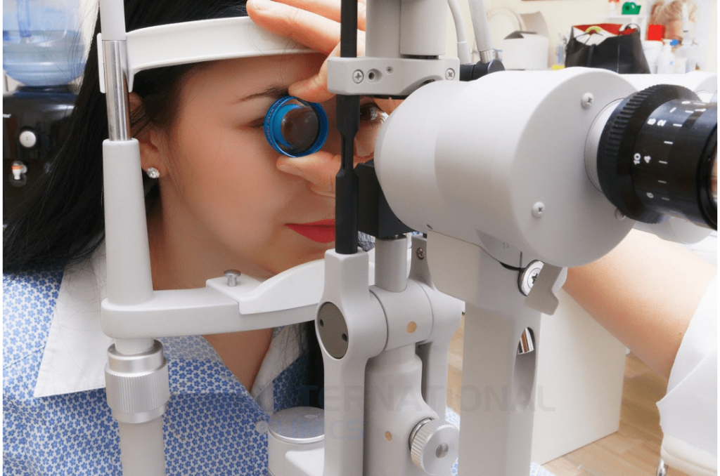
This is the process of dilating the eye using eye drops. Doctors carry out this process to enlarge the pupils and when shining light in the eyes. People describe it as an “old-fashioned” way to screen the eyes and diagnose eye conditions.
Dilation is used as the first step in different retinal examinations, but it’s incapable alone of diagnosing retinal conditions or diseases. It also produces some discomfort and may sting people.
Your pupils take about 20 to 30 minutes to fully open after dilation. Expectedly, you’ll experience blurry vision and minor sensitivity to light after dilation. The effect of dilation, however, usually disappears within a few hours.
Optomap Retinal Imaging
Retinal imaging has changed how doctors identify retinal diseases and monitor response to therapy. It provides a much broader picture of the eye by creating a series of images that the doctor can examine and compare.
Retinal imaging devices come in different technologies and functions. They include optical coherence tomography, fundus photography, angiography, and other types that can scan the retina and detect abnormalities.
Optomap retinal imaging is a unique, revolutionary technology that allows your doctor to get a high-resolution, ultra-widefield image of the retina. Retinal imaging and image analysis processes are achieved through digital computation inside the device.
It belongs to intelligent retinal imaging systems that help your doctor evaluate your retina and detect problems such as macular degeneration, diabetic retinopathy, retinal holes, and other problems. According to a study, Optomap imaging can identify additional retinal abnormalities compared to other methods of imaaging.
The retina is a thin layer that lines the back of the eye. It is located near the optic nerve, which is responsible for sight. The retina contains millions of light-sensitive cells (rods and cones). They are the neurons that are largely responsible for transmitting and regulating visual information.
Optomap Retinal Imaging vs Dilation: Benefits
Benefits of Dilation
Your doctor uses special eye drops to enlarge the pupils and force them to stay open. This process has a lot of benefits, such as:
- Easy and inexpensive
- Highly needed as part of a thorough eye exam
- Helps diagnose common eye problems, including glaucoma and age-related macular degeneration
Benefits of Optomap Retinal Imaging
Optomap retinal imaging or optos retinal imaging doesn’t involve eye dilation, but it’s not cheap and requires familiarity with the technology on the part of the doctor. However, it provides wide field retinal imaging and has a lot of advantages, including:
- Safe, painless, and doesn’t cause discomfort
- Provides the most comprehensive eye exam possible
- Fits all patients regardless of their age
- Instantaneous and does not affect your vision
- Can monitor changes in eye health over time and keeps records
- Produces images that can be exported to smartphones
- Helps detect many eye diseases, such as glaucoma, macular degeneration, and diabetic retinopathy
Optomap Retinal Imaging vs Dilation: Uses
Uses of Dilation
Your doctor can detect different problems during dilated eye exams. Below are some of these problems:
- Glaucoma: An experienced doctor can look for damage to the optic nerve and diagnose glaucoma during a dilated eye exam.
- Cataract: This condition occurs when your natural lens becomes cloudy.
- Diabetic retinopathy: When blood vessels of the eye leak or grow abnormally in the retina, your doctor may suspect diabetic retinopathy.
- Age-related macular degeneration: This condition occurs when the macula of the eye breaks down due to protein or pigment buildup.
Uses of Optomap Retinal Imaging
Besides being able to diagnose and monitor common eye diseases mentioned above, optos retinal imaging can also diagnose other eye conditions that are difficult to explore by the traditional dilated eye exams, such as:
- Retinal tear: This occurs as a result of pulling the transparent and jelly-like substance (vitreous) on the retina.
- Retinal detachment: The main cause of this condition is the presence of fluid under the retina. This happens when the fluid passes through the hole in the retina, which pushes the retina from the tissues underneath, causing a detachment of the retina.
- Epiretinal membrane: This is a superficial scar that resembles a sensitive tissue or a wrinkled membrane. It forms over the retina and attracts the retinal membrane to it, leading to blurred vision.
- Macular hole: This results from a small defect in the center of the retina at the back of the eye (the macula). The hole occurs due to abnormal pulling between the retina and the vitreous. It can also occur after an eye injury.
- Retinitis pigmentosa: Retinitis pigmentosa is a degenerative disease that affects the retina.
Optomap Retinal Imaging vs Dilation: Risks
Risks of Dilation
Dilation is not a pleasant examination for most people. The effect of eye dilation needs time to manifest, and the doctor needs 15 minutes to manually check the eye condition. The accuracy of eye dilation also highly depends on the doctor’s experience.
Eye drops can cause discomfort and, sometimes, serious side effects in case the patient was allergic to eye drops. Thus, it’s highly important for doctors to ask their patients about allergies before administering any eye drops.
Risks of Optomap Retinal Imaging
Optomap retinal imaging is very safe and only takes minutes to produce a highly detailed image of the retina. The FDA (food and drug administration) has approved its use to scan eye conditions according to reports.
Optomap retinal imaging is unlikely to cause any danger to the eye because it doesn’t touch the eye directly and relies only on low-powered laser wavelengths that scan simultaneously.
Difference Between Retinal Imaging with Injection & Without Injection
Retinal imaging is a simple, painless, and completely safe test. The imaging process commonly involves scanning the fundus of the eye by using a laser beam that passes through the layers of the retina. However, some retinal imaging methods involve injections. Optomap retinal imaging doesn’t involve injection or dilation of any sort.
- Retinal imaging without injections: This involves the placing of different eye drops on the eye and then using optical coherence tomography. However, this option may not bring accurate results.
- Retinal imaging with injection: This involves the injecting of sodium fluorescein into a vein in the arm or hand. The doctor dilates the pupil to facilitate the examination and detection of the retina. This test also depends on optical coherence tomography.
During the procedure, the patient sits in front of the retinal imaging device’s camera. The doctor takes several images of the retina and injects the patient again with sodium fluorescein upon the completion of the imaging process.
Treatments Methods of Retinal Diseases
After undergoing Optomap retinal imaging, the doctor will recognize the disease that affects the retina and set the stage for your treatment journey. There are many options through which retinal diseases can be treated. Here are some options:
- Laser therapy: Laser surgery can repair retinal tears or holes with great accuracy and in a safe way.
- Cryotherapy: This method tries to fixate the retina at a low temperature. The surgeon freezes the wall of the eye to treat a retinal tear. But the cold temperature must be intense and just enough to freeze the retina and treat the diseases.
- Air Injection: This method tries to fixate the retina with gas. Doctors use such a method when repairing certain types of retinal detachment.
- Eye surgery: Doctors perform eye surgeries to repair the detachment of the retina and other critical diseases.
- Draining the eye fluid: This process is also known as vitrectomy. The surgeon removes the “jelly-like” fluid that fills the inside of the eye. Then injects the eye with air, gas, or liquid to replace the fluid. This treatment is a good option for people with a retinal tear, diabetic retinopathy, macular hole, eye infection, eye trauma, or retinal detachment.
- Drug injection: The easiest way to treat retinal diseases is to inject a drug into the vitreous body of the eye. This method is effective in treating many diseases. It can be a good choice for people who have wet macular degeneration, diabetic retinopathy, or broken blood vessels in the eye.
Types of Eye Injections
Although injections are usually used in retinal imaging technologies other than Optomap retinal imaging, they also have a role to play in the treatment of various diseases that occur in the eye. Clearly, we’re not talking here about pigment injections, but about therapeutic injections.
In fact, many people prefer to use these injections, especially if they try to avoid surgery or if they have conditions that can be treated solely by injections. There are many types of eye injections, the most common include:
- Antibiotic injections: These can treat retinal infections, particularly bacterial infections that can affect sight.
- Cortisone injections: People can take these injections to treat uveitis and macular edema.
The Bottom Line
Optomap retinal imaging vs dilation! Which one is better than the other? obviously, Optomap retinal imaging is more useful and less risker than conventional eye dilation. However, it is certainly more expensive and not widely available.
International Clinics provides patients with the opportunity to undergo different retinal imaging tests, including traditional eye dilation and Optomap retinal imaging. Our doctors have successfully scanned the eyes of thousands of patients. You can reach us directly by using the Contact Us buttons below.
Frequently Asked Questions (FAQ)
Is Eye Dilation Painful?
Not exactly. Eye dilation is not a painful procedure, but you may feel a sting in your eye and certain level of discomfort. Most people tolerate these symptoms, and everything returns to normal after eye examination.
How Do I Get My Eyes Back to Normal After Dilation?
You must rest in your home and avoid going to work after getting eye dilation. Try also to rest in a dark room and wear sunglasses when go outside in the sun.
Why Do I Feel Sick After Getting My Eyes Dilated?
Unfortunately, some people experience unwanted symptoms after eye dilation because the body can absorb these drops and interact with them systemically. Some people may feel a spike in blood pressure, dizziness, and headache.
What Can Be Seen on Optomap?
Optomap device can help diagnose and see eye diseases and symptoms of systemic problems. This includes diabetic retinopathy, high blood pressure, macular degeneration, retinal holes, and retinal detachments.
Can Optomap Detect Cataracts?
Yes of course. Optomap retinal imaging can detect cataracts easily. However, doctors don’t rely only on Optomap retinal imaging to diagnose cataracts. There are other cheaper alternatives to detect cataracts.
Can Optomap Detect Glaucoma?
Yes of course. In fact, doctors can use Optomap retinal imaging to detect almost any type of glaucoma.
Can Optomap Detect Retinal Tear?
Optomap retinal imaging can detect retinal detachments without a problem; however, detecting retinal holes and tears might be a problematic due to limitations in the optics according to a study from 2009. Things are developed pretty much since then.
Read more: Glaucoma Treatment in Turkey: Surgery & Other Options
