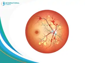
The macula is a very small area (5 millimeters in diameter) in the center of the retina at the back of your eye. However, despite being small, it may the macula could get damaged by multiple diseases or conditions, such as uveitic macular edema.
Macular edema may occur as a result of dehydration and enlargement of the macula. Patients in the advanced stages suffer from the accumulation of fluid, inflammation, swelling, or bleeding in the macula, which results eventually in macular degeneration.
The function of the retina is to transmit light through the optic nerve to the brain, so you can see clear images with details. The macula has important functions in this process as it helps your brain recognize people’s faces and makes you able to drive, read, and see in detail.
What Is Uveitic Macular Edema?
Uveitic macular edema is a retinal thickening in the macular area due to acute or chronic uveitis. Uveitis is the inflammation of the uvea or uveal tract in the middle of the eye, and it could lead to vision loss, especially among the youth, but doctors can treat it easily with medications.
Visual impairment is highly possible among people with uveitis and who also develop uveitic macular edema. The condition can lead to the accumulation of fluid within the retinal layers. Doctors always recommend early diagnosis and treatment to prevent long-term vision loss due to the condition. Uveitic macular edema treatment can include drugs and surgery.
Uveitic Macular Edema Traditional Treatments

Most doctors rely on medications to treat uveitic macular edema. They often prescribe steroidal medications along with other helpful drugs.
Steroids
Corticosteroids are still the standard option to deal with uveitic macular edema. Topical and nontopical steroids and available, and they include prednisolone, triamcinolone acetonide, methylprednisolone acetate, dexamethasone, and difluprednate.
Nonsteroidal Drugs
Nonsteroidal anti-inflammatory drugs are usually given as eye drops, but their efficacy is still controversial. Common names include bromfenac and nepafenac. It’s very important to take these eye drops cautiously to avoid their side effects.
Uveitic Macular Edema New Treatments
According to a study published in 2019, the effectiveness of steroid medications has been put into question by many researchers. The adverse effects of these medications and the tenacious nature of uveitic macular edema have led many doctors to start prescribing other promising medications, such as:
Immunomodulator Agents
These medications can interfere with DNA and RNA processes inside the cells to inhibit inflammation. Common names include methotrexate and azathioprine. Cyclosporine belongs to the T-cell inhibitors category, which is a group of immunomodulator agents.
Anti-Vascular Endothelial Growth Factor
Medications such as bevacizumab and ranibizumab are used by doctors to treat uveitic macular edema in a very limited number of cases because these medications don’t control inflammation and may even make it worse, but they’re still an alternative option to steroid medications.
Biological Agents
These agents have started to gain more attention in recent years, thanks to their effectiveness and limited side effects. Medications such as etanercept and infliximab could be used to treat uveitic macular edema, but it’s always recommended to consult a rheumatologist before doing that.
A study published in 2021 revealed that only half of the patients with uveitic macular edema respond to treatment. The study also stressed the futility of adopting a single treatment regimen for the condition because not all cases respond equally to medications.
Symptoms of Uveitic Macular Edema
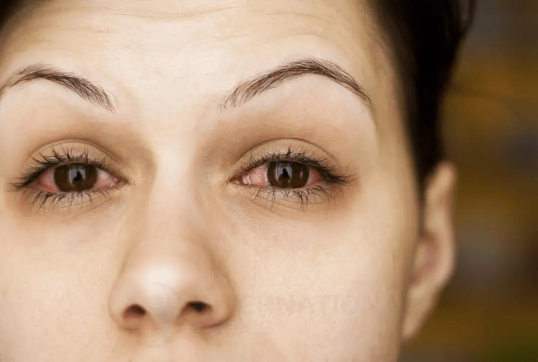
Before going to doctors, the patient could complain about several signs and symptoms that may indicate uveitic macular edema. Uveitis alone can cause eye pain and redness, along with other symptoms, such as light sensitivity and seeing floaters.
The main symptoms of macular edema include losing the ability to distinguish between colors and seeing blurred objects. Some patients may notice differences in the sizes of objects that they see. The patient may not feel any pain, but they could face difficulty seeing in low-light conditions.
Other Causes of Macular Edema
Uveitis is not the only cause or risk factor that may lead to macular edema. In fact, there are many more factors that can lead to the condition, including:
Aging
Aging is one of the risk factors that increase the risk of macular edema. In fact, the condition commonly affects people over the age of 50 who have macular degeneration.
Family History
If one of the parents suffers from a hereditary disease such as retinitis pigmentosa, their children may inherit the gene that causes the disease and increase their risk of having macular edema.
Blockage of Retinal Veins
Arteries are responsible for transporting oxygenated blood to various parts of the body, while veins are responsible for returning deoxygenated blood to the heart again.
Blockage may occur in the main vein that drains blood from the retinal tissue or in one of its associated branches. The possibility of this happening increases in older people and those who suffer from health conditions, such as diabetes mellitus, high blood pressure, or glaucoma.
Diabetes
Diabetes can cause many complications. The most common of which are macular degeneration and edema. High blood sugar expands the lenses spontaneously, which causes confusion.
In advanced cases, with the body’s sugar level remaining out of control, glaucoma may occur as a result of a high amount level of fluid in the eye. The pressure level rises in the eye, causing a gradual loss of vision.
Cataract Surgery
Cataract surgery helps remove lens clouding, which occurs as a result of protein accumulation in the lens area. Surgical intervention helps patients restore a large part of their natural vision. The duration of laser cataract surgery is between 15-30 minutes.
Cataract surgery tries to remove the cloudy lens and implant an artificial lens inside the eye. Doctors use an ultrasound device to break up the cloudy lens into small pieces, and then gently pull it out through a suction device. Complications after cataract surgery are rare, but they could include increased eye pressure, macular edema, and other retinal problems.
Other Causes
Macular edema may result from other inflammatory diseases, such as sarcoidosis. Also, the condition may develop due to toxicity, trauma, or unknown causes.
How to Diagnose Macular Edema?
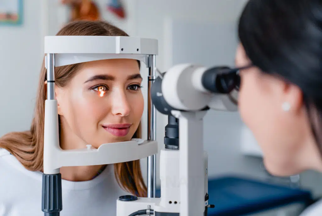
Thanks to its sensitive features, optical coherence tomography is the standard tool to diagnose macular thickening. However, there are many other ways to diagnose macular edema. We can review them in detail as follows:
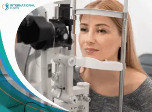
Ophthalmoscopy
Ophthalmoscopy is one of the important techniques that detect macular problems and other eye diseases. The doctor looks through the pupil of the eye and identifies any problems in the retinal blood vessels, the optic nerve, or the fundus of the eye. The duration of this examination is between five and ten minutes. Both direct and indirect ophthalmoscopy can be helpful.
Fluorescein Angiography
Fluorescent angiography is among the important tests for diagnosing macular edema and other eye diseases. The history of this examination dates back more than half a century. The doctor injects a contrast dye into the central vein to identify any problems in the blood flow inside the eye.
It is prohibited to use this method to diagnose macular edema in pregnant women or those who are allergic or have problems in their arteries, heart, or liver.
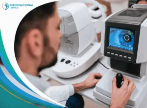
Optical Coherence Tomography
Optical coherence tomography (OCT) is one of the primary methods for diagnosing macular edema as it helps obtain accurate detailed images of the retina to identify appropriate measures for treating macular degeneration. Doctors have extensively relied on this device since 2005. It made a remarkable revolution in the field of eye diagnostics.
More About the Macula
Below is more information on the anatomy and function of the macula:
- The macula contains a cavity known as the fovea. Oxygen reaches this area through extended blood vessels that pass through Bruch’s membrane, an intermediate layer between the fovea and the choroid.
- The middle of the fovea (fovea centralis) contains conical cells that are very sensitive to colors. When you focus your vision, the light rays coming from the lens fall on the fovea centralis, which helps you see clear images. Thus, this part is responsible for the sharpness of vision.
- The number of cone cells is 4.5 million, while the number of rod cells is approximately 91 million in each eye. Despite the low sensitivity of cone cells to light compared to rod cells, cone cells help distinguish colors and perceive detailed images because their response rate is higher than that of rod cells.
- The visual acuity in the Fovea centralis is 20/20. The fovea centralis is located in the opposite part of the eye lens and is responsible for distinguishing colors and seeing the smallest details. Studies indicate that the fovea has the highest sensitivity to light in the whole eye.
- The fovea centralis is surrounded by two maculas within the retinal area. The first macula has a diameter of 1.25 millimeters and contains five layers of retinal ganglion cell cells. The second macula surrounds the first macula and has a diameter of 2.75 millimeters. It consists of five layers of retinal neuron cells. The density of cone cells in fovea centralis is 12 cells per 100 micrometers. There is also a third macula that surrounds the previous two.
- The information transmitted from the macula to the brain represents 50% of the total images that the brain translates and understands, while the other 50% represents the information transmitted through other areas of the retina.
The Bottom Line
For more than 5 decades, doctors have used to deal with uveitic macular edema using eye drops containing steroids. However, recent studies and investigations revealed a possible role for other medications, such as immunomodulator and biological agents, in treating the condition.
optical coherence tomography remains the single most used tool to diagnose uveitic macular edema and other retinal diseases. The progressive advent of technology led to better accuracy in diagnosing retinal conditions and diseases.
International Clinics provides reliable diagnoses and treatment for uveitic macular edema patients. You can reach us directly by using the Contact Us buttons on our website.
Frequently Asked Questions (FAQ)
What Is the Best Treatment for Macular Edema?
There is no specific best treatment for macular edema because each case responds differently to medications, and there is no one eye drop that can solve all cases. However, anti-VEGF drugs are known for being very effective, at least in some cases.
Does Macular Edema Go Away?
Macular edema doesn’t go away on its own. But, in rare cases, the condition can improve and seemingly go away without external intervention. In any case, macular edema can lead to vision loss and even blindness; hence, it’s very important to never overlook the condition.
Which Vitamin Causes Macular Edema?
Vitamins are important for bodily processes, but taking some vitamins in large amounts could lead to problems. Vitamin B3 (niacin) is suspected to cause macular edema when taken at very high doses.
Can Glasses Help Macular Edema?
Macular edema isn’t a condition that glasses can treat. Macular conditions in general are complicated and may require invasive surgeries to cure them.
What Is Uveitic Cataract?
Uveitis could result in cataracts in some cases due to corticosteroid use and chronic inflammation, leading to “uveitic cataracts”. Despite being a challenging condition, doctors can treat uveitic cataracts through different methods.
What Is Uveitic Glaucoma?
Uveitis could result in glaucoma in some cases too due to chronic inflammation, leading to “uveitic glaucoma”. Glaucoma can be managed by using eye drops and surgery.
Do People With Uveitis Go Blind?
Uveitis can lead to blindness and vision impairment. However, early diagnosis and treatment can decrease the chance of blindness and treat the case for good.
Is Uveitic Macular Edema an Orphan Disease?
Macular edema in general isn’t an orphan disease. Diabetic macular edema appears in 746,000 adults over 40 years of age in the United States. However, the prevalence of uveitic macular edema seems much lower compared to diabetic macular edema.
Read also: Glaucoma Treatment in Turkey