The commercial systems of optical coherence tomography (OCT) are diverse and depend on different methods to detect signals from the retina. These methods include time-domain OCT, spectral-domain OCT, and swept-source OCT. Stratus optical coherence tomography commonly belongs to the time-domain category.
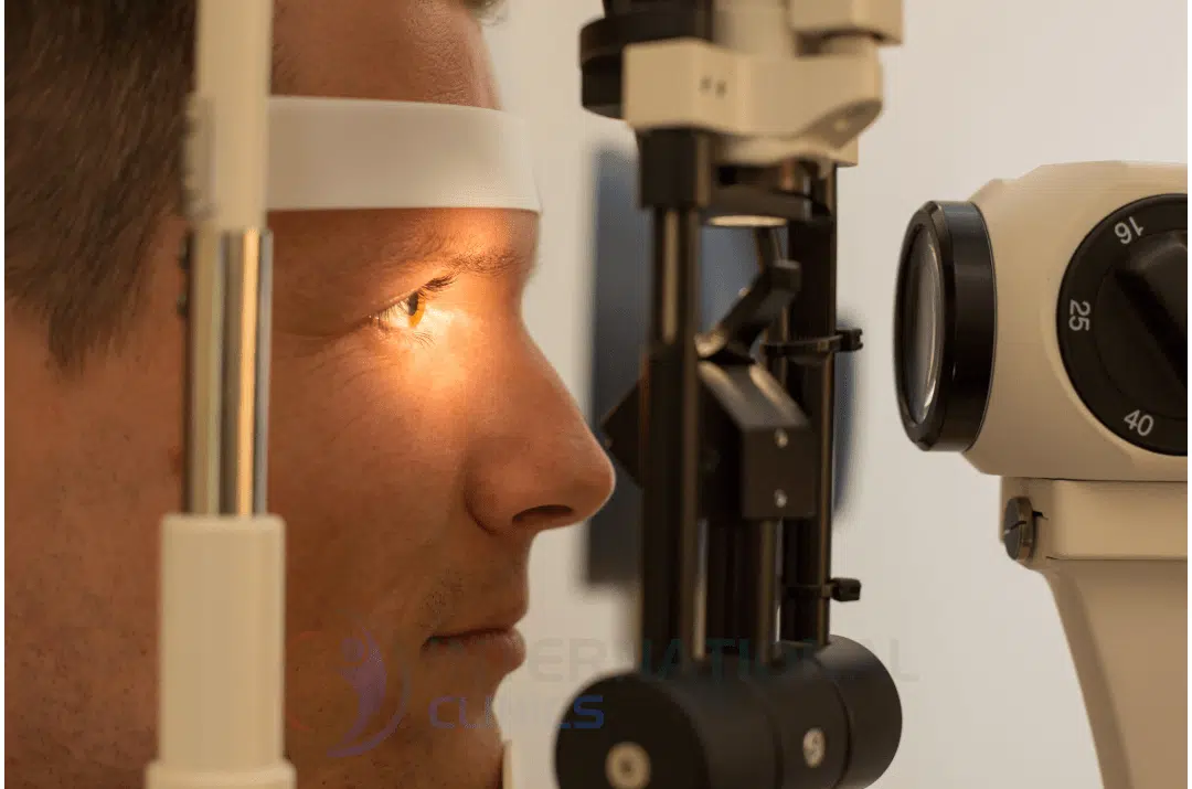
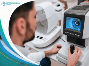
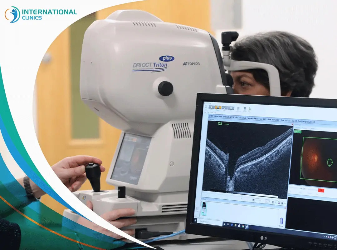
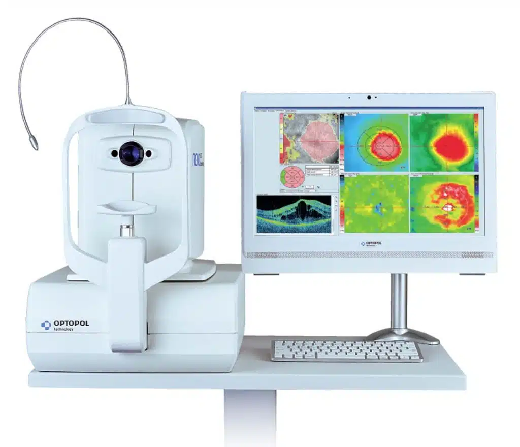
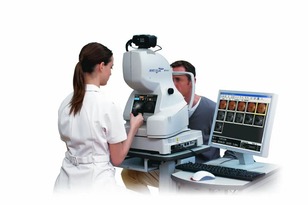
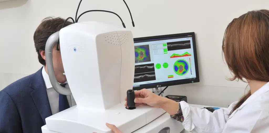
Retinal tomography or optical coherence tomography (OCT) is generally used to scan the retina (back of the eye) and get high-resolution cross-sectional images. This technique utilizes light waves to take precise and detailed images of the retina and detect impairments and defects.
It also helps examine the anterior and posterior parts of the eye to arrive at the right diagnosis and treatment options in later stages. This technique is similar to computed tomography and magnetic resonance imaging. People commonly seek an OCT eye scan to investigate different eye diseases, such as glaucoma.
Stratus Optical Coherence Tomography: History
Optical coherence tomography was first developed in 1991 at the Massachusetts Institute of Technology. However, after just 4 years of its first inception, the device was ready to get into the market, particularly after the German manufacturer Carl Zeiss acquired the device.
Optical coherence tomography devices have gone through persistent evolution, leading eventually to the creation of Stratus optical coherence tomography (Stratus OCT), which succeeded the first generations of optical coherence tomography and becomes a standard instrument in ophthalmology.
The demand for Stratus OCT is increasing rapidly as many studies confirmed its efficacy and many doctors have mastered its use. In fact, more than 6,000 units of Stratus optical coherence tomography machines were sold by 2006. Popular brands include Zeiss.
Stratus Optical Coherence Tomography: Uses
Doctors use Stratus OCT to accurately diagnose many diseases and obtain ocular images after dilating the eye pupils.
Diagnose Glaucoma
Glaucoma is a common eye condition in which the optic nerve is damaged as a result of high intraocular pressure. This leads to vision impairments. It mostly affects people over the age of sixty and to a lesser extent younger people.
Often, the symptoms of this condition don’t appear immediately. As the condition progresses, symptoms start to appear. One of the important tests to diagnose this disease is Stratus OCT.
Symptoms of glaucoma include eye redness, severe headache, squinting, eye pain, vomiting, nausea, and blurred vision. If left untreated, this may lead to vision loss. The main risk factors that increase the chances of developing glaucoma are the following:
- Taking corticosteroids for too long
- High intraocular pressure
- Injury to the eye or undergoing surgery in the eye
- Longsightedness or nearsightedness
- Having a family member who previously developed the disease
- Being Latin or African descent
- Having diseases, such as heart disease, tuberculosis, sickle cell anemia, and high blood pressure
- Being over sixty years old
- Corneal thinning
Diagnose Age-related macular degeneration
Age-related macular degeneration can lead to complete loss of central vision. This condition is often described as a “progressive” disease and usually occurs in the elderly. However, doctors can treat some forms of age-related macular degeneration with medication and surgery.
Diagnose Diabetes-related retinopathy
Diabetes-related retinopathy occurs as a result of damage to the small blood vessels of the eye. This condition can lead to glaucoma and, possibly, complete vision loss, especially when the entire retina detaches from the back of the eye. Luckily, doctors can suggest a surgery called “vitrectomy” to treat this condition.
Diagnose Progressive Retinal Atrophy
Progressive retinal atrophy is among the genetic diseases that cause a gradual loss of vision. Most cases have choroidal membrane atrophy. In some cases, this occurs parallel to atrophy in other areas of the body, not just the eye, as is the case of some syndromes. This disease is among the rare diseases and can be diagnosed by optical coherence tomography.
Diagnose Retinoblastoma
Retinoblastoma is a malignant type of cancer that affects the retina. It mostly occurs in young children, and to a lesser extent in adults. The retina consists of nerve tissues that transmit light from the front part of the eye.
The signals reach the brain via the optic nerve and the brain translates them into images. Optical coherence tomography helps diagnose the tumor. The symptoms of retinoblastoma include eye swelling and redness, asymmetrical eyes, and white-colored pupil (the center of the eye).
Retinoblastoma originates from the abnormal growth of cells that make up the retina. The tumor can spread to other places in the body, such as the spine and brain. In the majority of cases, the cause of the genetic mutation that leads to the emergence of the tumor remains unclear.
Diagnose Other Eye Conditions
Doctors could use Stratus optical coherence tomography to diagnose conditions such as cystoid macular edema, macular hole, macular pucker, and cone and cone-rod dystrophies.
Prepare for Refractive Surgery
Candidates who undergo refractive surgery, such as LASIK, may need to be examined by Stratus OCT. After knowing the right diagnosis, they can undergo surgery to correct longsightedness, myopia, or astigmatism, or to treat keratoconus disorder, which usually requires laser therapy to reshape the curvature of the cornea and correct the vision.
Stratus Optical Coherence Tomography: Features & Protocols
In the early days of the OCT devices, the level of modernity and development was primitive and required more innovation. In recent times, however, the device has become more advanced as many technical changes and advancements have occurred.
The Stratus optical coherence tomography utilizes various scanning programs or protocols, such as the macular thickness map (MTM), the fast macular thickness map (FMTM), line scan, and cross-hair scan protocols. These protocols enable many features, such as:
- Easier diagnosis of macular edema and retinal disorders.
- Better visualization of blood circulation in the retina.
- Examination of the retina through the device does not require much time or effort.
- Easier early diagnosis of many eye disorders.
Stratus Optical Coherence Tomography: The Procedure
The patient places his head in a particular chamber, and the technician adjusts the imaging device and instructs the patient to look at a target area in the device. The pictures are taken, and eye drops can be used to dilate the pupil and obtain pictures of specific areas, but usually, nothing will touch your eyes.
The examination takes only five minutes and there are no special preparation steps that you need to follow before undergoing the procedure. All you need is to focus your eyes on a green object within the machine to help the laser inside the machine to create pictures of the layers of your retina and optic nerve.
Stratus Optical Coherence Tomography: Drawbacks
Stratus OCT technology is no more the only game in town. In fact, the new types of spectral-domain OCT devices are gaining more momentum as they provide better and faster results compared to the traditional time-domain OCT, such as Stratus OCT.
Cirrus OCT is a spectral domain OCT that can provide better image resolution with higher sensitivity and speed. This device has recently become available for purchase by many clinics around the world. What makes this device special is its ability to generate better-defined retinal images approximately 70 times faster than Stratus OCT technology. However, some studies found no significant difference between the two types.
More About Optical Coherence Tomography (OCT)
Here are some quick general facts about optical coherence tomography:
- The optical coherence tomography device uses light waves to scan structures of the eye. The production of the device has begun in 1994, but it was not used extensively. Indeed, its presence was limited to large treatment centers. However, the demand for using the device exploded in 2005 as its cost becomes cheaper. Since then, there is no center or private clinic that does not use an OCT retinal scan.
- The demand for purchasing an optical coherence tomography machine is still growing because of its remarkable ability to diagnose many eye diseases that were difficult for doctors to diagnose accurately in the past.
- Light waves used by OCT can penetrate various areas of the retina and obtain very precise details of the internal structures.
- The performance of optical coherence tomography devices has also improved in recent years, which further enhanced monitoring, diagnosing, and following up on the effect of the treatments given to patients who suffer from retinal and macular eye diseases.
- The device helps obtain detailed images of the retinal nerve fibers, which is very important for patients who have glaucoma and diabetic retinopathy.
The Bottom Line
Stratus optical coherence tomography (Stratus OCT) has become a staple technology to diagnose different eye conditions, including glaucoma and diabetes retinopathy. The features and protocols of the device make it unbeatable in terms of value and significance.
Here at International Clinics, our doctors use different kinds of optical coherence tomography devices to scan the eyes of patients and detect abnormalities that otherwise couldn’t be detected through other means.
Frequently Asked Questions (FAQ)
What Is Stratus OCT?
Stratus optical coherence tomography (Stratus OCT) is a non-invasive imaging technology that uses light waves to capture high-resolution, two- and three-dimensional images of the retina after dilating the eye pupils.
What Is the Cost of an OCT Scan?
Depending on the clinic or hospital, the cost may range between 50-1000 US dollars. The type of device, consultation fees, and reputation of the hospital are some of the factors that play a role in determining the cost of the test.
Do I Have to Pay for an OCT Scan?
Mostly yes. The casual eye test doesn’t include OCT. Hence, you may need to pay additional fees if your doctor finds it important to get an OCT scan.
How Often Should You Have an OCT Eye Test?
If you’re at risk of developing eye diseases, your doctor may recommend you undergo an OCT scan, especially if you’re older than 25 years. You may have it as a part of your annual eye screening.
Can an OCT Scan Detect a Brain Tumor?
Yes of course. An OCT is used by doctors and researchers to detect the spread of certain brain tumors. However, it’s not the main method to detect brain tumors.
Can an OCT Scan Detect Diabetes?
An OCT can detect damaged blood vessels in the retina, which may signal the presence of diabetic retinopathy, which is a complication of diabetes.
Can an OCT Scan Detect Retinal Detachment?
Yes absolutely. An OCT can be a great method to diagnose retinal detachment and other problems that damage the retina.
Read more: Eye Surgery Preparation