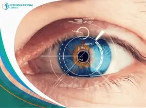
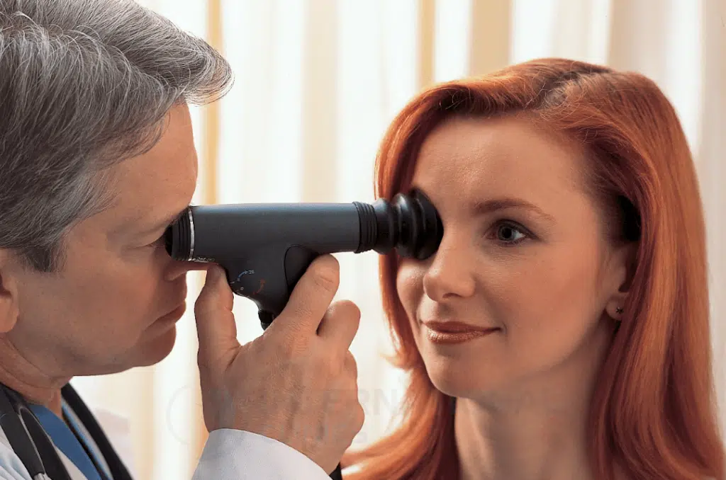
Ophthalmoscopy (funduscopy) involves the use of an ophthalmoscope to assess the back parts of the eye (the retina). This examination can identify the condition of the optic nerve, the inner eye cavity, and blood vessels.
Ophthalmoscopy is very important to determine the extent and severity of eye conditions, especially for those who suffer from glaucoma, diabetes, or high blood pressure.
There are many reasons that prompt the doctor to conduct ophthalmoscopy, including the increased risk of developing eye diseases for people who have a family history of these diseases, including glaucoma, squint, or others.
Ophthalmoscopy divides essentially into 3 types: direct, indirect, and slit-lamp ophthalmoscopy. In this article, we will focus mainly on the indirect ophthalmoscope and how it differs from other types.
What Is Indirect Ophthalmoscopy?
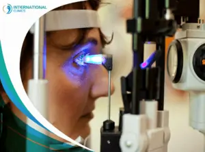
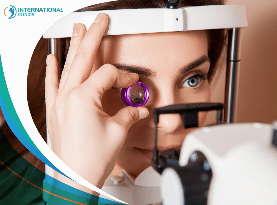
An indirect ophthalmoscope or binocular indirect ophthalmoscopy (BIO) helps the doctor view the retina with a moving device. They use a handheld condensing lens to create a dynamic observation. The doctor wears the device around the head and directs the binocular lenses in front of the eyes.
The indirect ophthalmoscope consists of a light source, a binocular lens with mirrors, and a headband. The Indirect ophthalmoscope helps your doctor get a much better view of the entire retina and see the front portions, which are hard to view with a direct ophthalmoscope. They can also get a better look at the back of the eye.
Why Choose Indirect Ophthalmoscopy?
According to a study, indirect ophthalmoscopy has great advantages that make doctors more willing to use it instead of direct ophthalmoscopy. Below are some of these advantages:
- Allows for a wider field of view and depth perception
- Penetrates moderate cataracts, thanks to its brighter light source
- Helps examine the anterior retina with scleral indentation
- Stable and isn’t affected by the refractive state of the patient’s eye
- Can be used in a sterilized operating room
Steps of Indirect Ophthalmoscopy
The indirect ophthalmoscopy is a precise examination that requires experience to perform. The examination only takes about 5 to 10 minutes. Often, the steps of this examination include the following:
Dilate the Eyes
First, your doctor will dilate your eye by using special eye drops. This will make it easier for the doctor to work on your eyes.
Set the Right Position
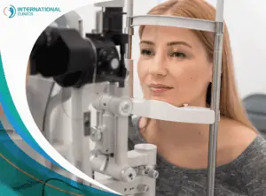
Proper positioning of the patient is very important for indirect ophthalmoscopy. You must lay flat in a reclining chair with enough space to allow for free head movement.
Choose Lens & Minimize Distortion
Your doctor will choose the right lens that has the optimal magnification and a dynamic field of view. They also will minimize lens distortion by moving the lens in and out in specific directions.
Adjust the Headband
Your doctor will adjust the headband that contains the lens to ensure stability. They also will adjust the light beam and light aperture.
Depress the Sclera
Scleral depression is necessary to get a dynamic viewing of the retina, especially if you have retinal tears or detachments. Your doctor could use a topical anesthetic to make you feel more comfortable during the examination.
Things to Consider Before Indirect Ophthalmoscopy
Many sources describe the indirect ophthalmoscope as one of the challenging pieces of equipment to master. Before undergoing indirect ophthalmoscopy, you must consider the following things:
- You must meet your doctor to discuss your family history and ensure that you don’t suffer from diseases that prevent the use of eye drops or increase intraocular pressure.
- The patient must use sunglasses upon completion of the procedure to alleviate blurry vision, which results from using an eye drop that expands the pupils.
- It is better for people with jobs that require clear vision to have a day off until their vision returns to normal.
- There are some medications that can interact in some way with eye drops used during the examination. Therefore, you must speak with the doctor and inform them of all the medications you’re currently taking or were taking in the recent past.
Other Types of Ophthalmoscopy
Indirect ophthalmoscopy is one type of ophthalmoscopy. Depending on your case, your doctor may find it better to choose another type of ophthalmoscopy, such as:
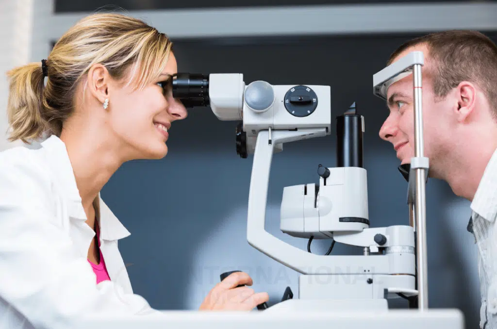
Direct Ophthalmoscopy
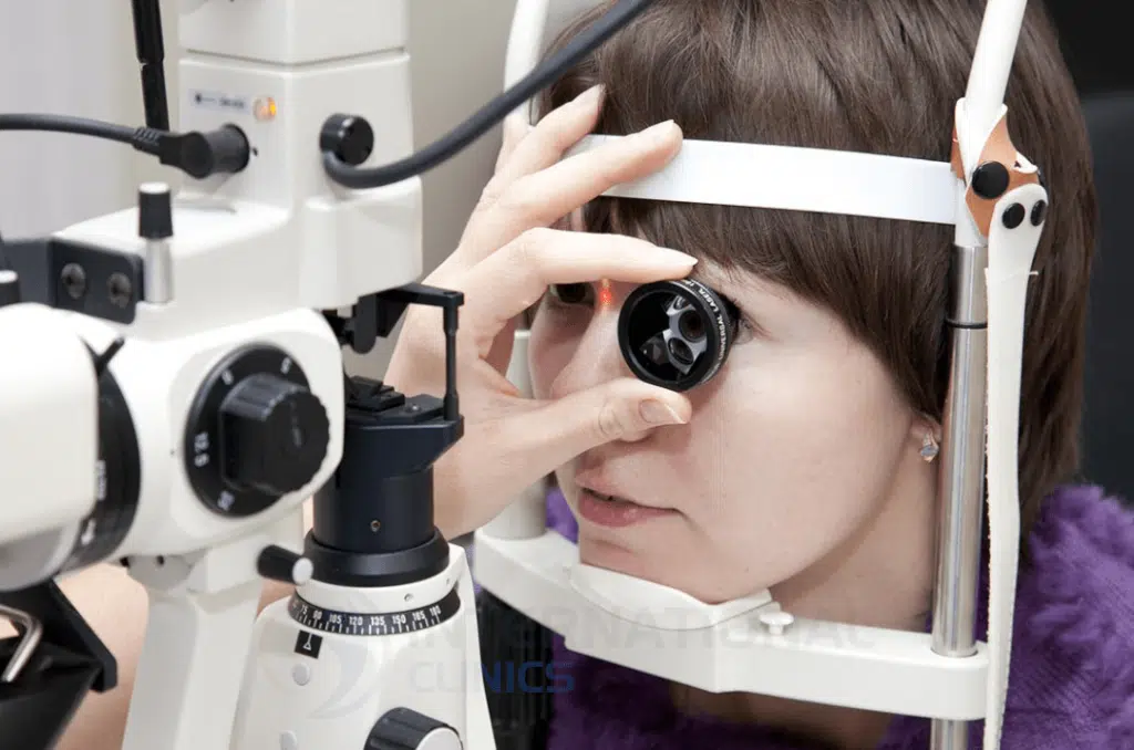
The doctor uses an ophthalmoscope in a dark room to get a better look at the retina. The patient sits facing the doctor, who examines the fundus of the eye with an ophthalmoscope that emits a beam of light into the pupil. To facilitate looking at the back of the eye, the doctor asks his patient to stare directly ahead and keep the head steady.
Slit-Lamp Ophthalmoscopy
This procedure requires you to look at the light produced by the slit lamp, which has a special base to rest your chin on. The doctor asks you to move your eyes in more than one direction so that they can examine the entire fundus of the eye using a microscope that enlarges the front of the eye.
Extended Ophthalmoscopy
For certain health policies, extended ophthalmoscopy becomes a necessity if a complete view of the back of the eye is needed but can’t be achieved by the standard direct ophthalmoscope, the indirect ophthalmoscope, or both.
Extended ophthalmoscopy can be a very helpful method to screen a wide range of conditions that affect the back portion of the eye. In any case, extended ophthalmoscopy is commonly used as an ophthalmoscopy code that healthcare professionals use to document a comprehensive direct or indirect ophthalmoscopy.
The results of Indirect Ophthalmoscopy
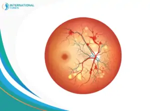
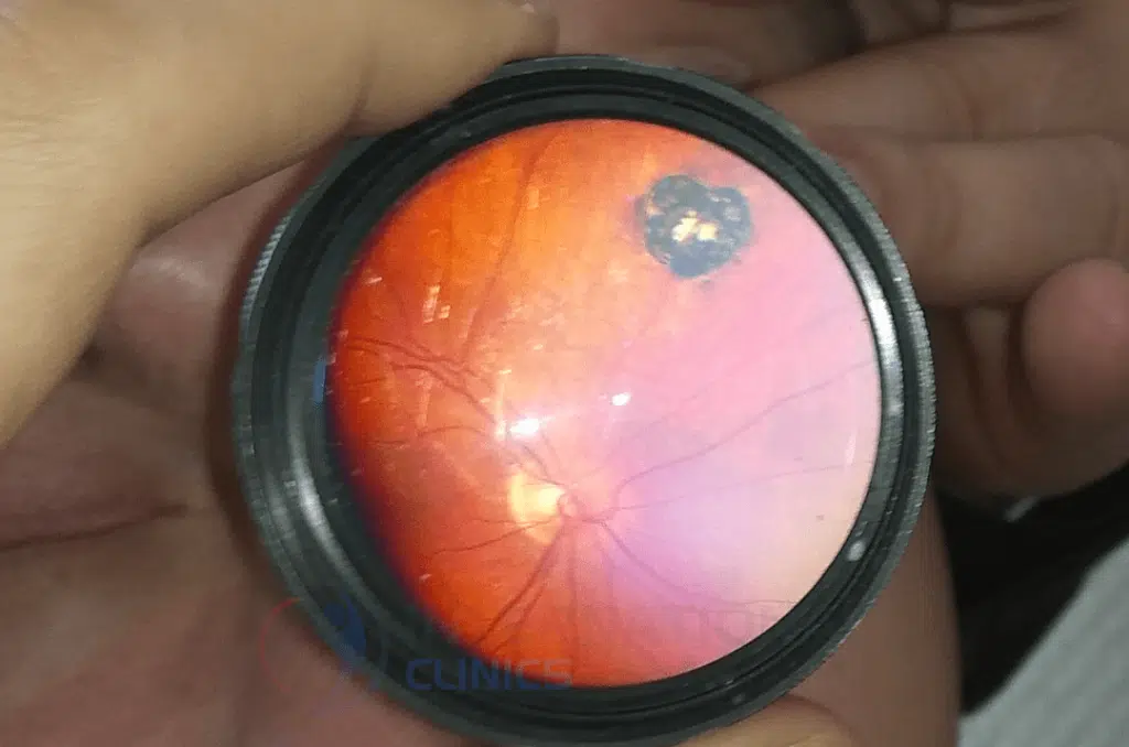
Indirect ophthalmoscopy is accurate enough to check the condition of the retina and optic disc. The doctor starts to suspect a problem in the retina if they notice spots or if the retina is swollen. Depending on the experience of the doctor, ophthalmoscopy can help detect conditions such as:
- Optic nerve problems
- Hypertension (High blood pressure)
- Different types of glaucoma
- Age-related macular degeneration
- Retinal detachment and tears
- Retinitis (viral inflammation of the retina)
- Melanoma of the eye
- Diabetes
Indirect Ophthalmoscopy for Infants
In infants, the vision is still premature and needs time to develop normally. However, any problem that may affect the vision must be recognized from day one. It will be easier to deal with conditions in their early days. Delays can complicate treatments.
Indirect ophthalmoscopy is often the right choice to screen eye conditions in infants. However, doctors commonly face challenges when performing this examination in infants, especially when it comes to dilating the eyes.
After six months of age, the newborn must undergo a fundus examination to determine if the vision is normal or not, especially if the child was born earlier than previously anticipated. Also, an eye examination is important if there is a family history of eye conditions or if the mother was exposed to health problems during her pregnancy.
At preschool age, the child should undergo another examination of the eye fundus to ensure that there is no vision problem that may become an obstacle to academic progress.
One of the most common eye conditions in children is nearsightedness, which prompts the doctor to check the child’s vision again to determine the sizes of the eyeglasses that fit their needs. If the child has astigmatism, then a comprehensive eye examination becomes very crucial.
Reasons to Perform Indirect Ophthalmoscopy in Infants
Examination of the eye fundus leads to early detection of problems before they occur, which makes treatment more efficient. Here is why infants should undergo ophthalmoscopy:
- If the infant is expected to have a vision problem that requires eye surgery.
- Children with special medical conditions- such as arthritis, Down syndrome, or neurofibromatosis- are more likely to develop future vision problems.
- Children with congenital glaucoma as a result of genetic errors or exposure to problems during pregnancy.
- Having a family history of myopia.
- Significant low birth weight.
- The mother’s exposure to a viral infection during pregnancy, or her infection with HIV, venereal diseases, and German measles. Also, if the mother is a smoker or addicted to different types of drugs.
- Having a difficult birth may increase the child’s risk of developing eye conditions.
Risks of Indirect ophthalmoscopy
Indirect ophthalmoscopy is uncomfortable for both the doctor and the patient. Often, people report seeing images after the light has been turned off. Thankfully, these images disappear spontaneously after blinking the eyes several times.
Indirect ophthalmoscopy is 90% to 95% accurate and rarely can cause any damage to the eye. However, some cases may develop problems related to the eye drops used during the examination, such as:
- Vomiting or dry mouth
- Flushing
- Nausea or dizziness
- Developing certain types of glaucoma
The Bottom Line
Indirect ophthalmoscopy or binocular indirect ophthalmoscopy can screen many eye diseases during a routine eye exam. This type of ophthalmoscopy requires the doctor to wear a headband that contains mirrors and lenses. Indirect ophthalmoscopy has many advantages that challenge the use of conventional, direct ophthalmoscopy.
Doctors at International Clinics use both direct and indirect ophthalmoscopy to check the conditions that affect the eye. You can reach us directly by using the Contact Us buttons below.
Frequently Asked Questions (FAQ)
What Is Indirect Fundoscopy?
Indirect fundoscopy is another name for indirect ophthalmoscopy, which is a great method to screen for retinal diseases and get a better view of the fundus. Doctors can check the presence of many eye conditions using indirect fundoscopy, such as retinal tears and detachment.
What Can an Ophthalmoscope Detect?
Doctors use the ophthalmoscope to detect different eye conditions, such as glaucoma. An ophthalmoscope can be very helpful also in detecting blood vessel problems inside the eye.
Who Invented Indirect Ophthalmoscope?
Indirect ophthalmoscopy was first introduced by Christian Ruete in 1852 after modifying the original design of the direct ophthalmoscope developed by Hermann von Helmholtz. The goal was to allow for a wider view of the fundus.
Is Direct or Indirect Ophthalmoscopy Better?
Both types can produce great and accurate results. However, indirect ophthalmoscopy provides a wider field of view and other advantages that surpass those of traditional, direct ophthalmoscopy.
What Structures Can Be Seen Through the Ophthalmoscope?
A doctor can see the fundus of the eye (the back part of the eye) by using an ophthalmoscope. Structures include the optic disc, choroid, retina, and blood vessels.
Which Direct Ophthalmoscope Is Best?
Many direct ophthalmoscope brands are available, such as WelchAllyn, Keeler, Heine, and Rudolf Riester. However, according to reviews and reports, Heineophthalmoscopes are the best, especially the Heine mini 3000 ophthalmoscopes and Heine beta 200 ophthalmoscopes. Ophthalmoscope Tarkov is a fictional ophthalmoscope that appears in a game called “Escape from Tarkov”.
How Much Is an Ophthalmoscope?
Despite being sophisticated devices, ophthalmoscopes are actually cheap and easy to get. You can get an ophthalmoscope for only 60-100 US dollars. However, the top-tier devices are sold for more than 1000 US dollars. Some can reach up to 9000 US dollars.
Read more: The Best 4 Eye Pressure Measurement Methods