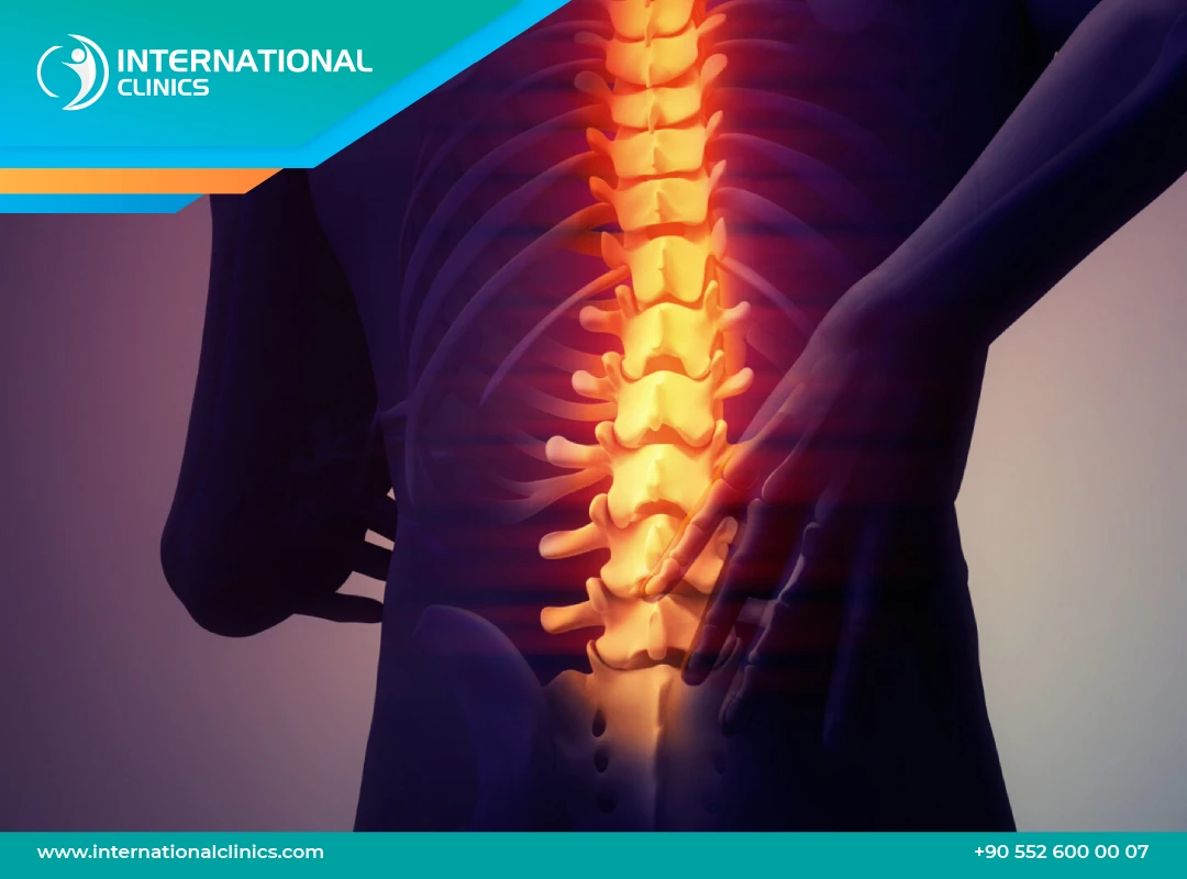
Spinal cord tumor surgery is a delicate operation to remove a tumor, entirely or as much as is safe, from inside or around the spinal canal. Its goal is to relieve pressure on the spinal cord and nerve roots, obtain tissue to identify the tumor type, and possibly ease pain, weakness or altered sensation.
It is done under general anesthesia. The surgeon reaches the canal from the back by removing part of a vertebra's bony arch, then separates the tumor from nerve tissue using a microscope and fine instruments, often with electrical nerve monitoring. The spine may be stabilized if needed, and the tissue is examined under the microscope.
Surgery usually takes several hours, followed by a hospital stay of several days. Physiotherapy starts early when possible, and rehabilitation may last weeks to months. Recovery depends on nerve function before surgery and the tumor type, and the final result is assessed with imaging and examination over several months; further treatment may be needed.
The final cost is set after a medical consultant reviews your case and photos.
Our medical consultants review all requests regularly, every three hours.
Get a free consultation and answers to all your questions by completing the form below
It may relieve pressure on the spinal cord and nerves, allows the tumor type to be identified precisely from the sample, and may limit worsening of symptoms. Results vary with the tumor type, its location and the patient's condition before surgery.
It is suggested for people whose MRI shows a tumor pressing on the spinal cord or nerves, or who have increasing pain, weakness or numbness. The surgeon and the oncology team decide whether it is suitable after reviewing the images and the patient's general health.
Blood tests, MRI and sometimes other investigations are done, and the anesthesiologist assesses your health. Tell the team about all your medicines, and never stop any of them on your own, especially blood thinners; the doctor decides what to stop and when.
Under general anesthesia, an incision is made in the back at the level of the tumor and part of the bone is removed to expose the spinal canal. The tumor is then removed with a microscope and fine instruments, often with nerve monitoring, the layers are closed, and the spine may be stabilized when needed.
Patients stay in hospital for several days and may start moving and physiotherapy early. Rehabilitation can last from weeks to months, pain relief is prescribed, and you are given instructions to protect your back and avoid heavy lifting.
Pain may ease early, while strength and sensation improve gradually and may not fully return. The outcome depends on the tumor type and how much was removed, and regular imaging follow-up is needed because some tumors can return or require further treatment.
They include bleeding, infection, cerebrospinal fluid leakage, leg blood clots, and nerve injury that may cause weakness, numbness or bladder and bowel control problems; complete removal of the tumor may not be possible. The surgeon explains and assesses these risks for each patient.
Yes, a vertical scar remains in the middle of the back, and its length depends on how many vertebrae are involved. It improves over time and is usually covered by clothing.
A hospital stay of several days is usually needed, followed by additional time for monitoring and wound checks before travel, which may extend to two weeks or more. Your treating doctor determines the right timing based on your condition and means of travel.
This may be necessary depending on the microscopic examination of the tumor, for example radiotherapy, drug treatment or imaging follow-up. The treating team will discuss the plan with you once the result is available.