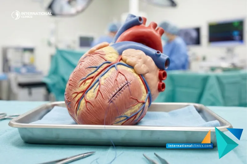
Tetralogy of Fallot surgery is an open-heart operation for a congenital defect with four parts: a hole between the ventricles, a narrowed outlet from the right ventricle to the lungs, an aorta sitting over the hole, and a thickened right ventricle. It aims to improve blood flow to the lungs and reduce bluish skin and low oxygen levels, and the treating team decides the timing for each child.
It is done under general anaesthesia with a heart-lung machine. The surgeon closes the hole with a patch and widens the right ventricle's outlet by removing the muscle causing the narrowing. A second patch or a pulmonary valve repair may be needed.
The operation usually takes several hours, followed by a few days in intensive care and about one to two weeks in hospital. Colour and stamina improve gradually, but the final result is judged through regular follow-up over many years. Some patients later need pulmonary valve treatment, and no outcome can be guaranteed.
The final cost is set after a medical consultant reviews your case and photos.
Our medical consultants review all requests regularly, every three hours.
Get a free consultation and answers to all your questions by completing the form below
It may improve blood flow to the lungs, raise oxygen levels and reduce blue spells, helping the child grow and be more active. This depends on each child's condition, and the decision rests with the treating team.
Children diagnosed with tetralogy of Fallot after assessment with an echocardiogram, and sometimes a cardiac catheterisation or CT scan. The heart surgeon and the paediatric cardiologist decide the best timing according to symptoms and heart anatomy.
The child has heart and blood tests, and the team monitors feeding, weight and oxygen levels. No medication is given or stopped except on the doctor's instructions, and the team explains the stages of the operation and aftercare to the parents.
Under general anaesthesia and with a heart-lung machine, the surgeon opens the chest, closes the hole between the ventricles with a patch and widens the right ventricle's outlet. The pulmonary valve may also be treated when needed.
The child spends a few days in intensive care and then on the cardiac ward until breathing, heartbeat and feeding are stable. Energy improves gradually over several weeks, and the doctor says when normal activities can resume.
Improvement in colour and oxygen levels is often noticed after surgery, but a full assessment needs long-term follow-up. Further procedures on the pulmonary valve or the right ventricle's outlet may be needed later, depending on the case.
They include bleeding, infection, heart rhythm disturbances, a leaking pulmonary valve and a small residual hole, and some can be serious. The surgical team explains the risks expected in your child's case.
Yes, a vertical scar usually remains in the middle of the chest. It fades with time but usually does not disappear completely.
Yes. The patient needs regular follow-up with a cardiologist, with echocardiography and ECG, often for life. The doctor may recommend preventive measures before some dental treatments and operations.
This depends on the child's stability, and it is often not before two to four weeks after the operation, sometimes later. The treating doctor confirms when and how to travel.